Mixed Cranial Nerve Schwannoma: A Rare entity Recognition and Key Imaging Features
Authors
Keywords:
- Array,
- Array,
- Array,
- Array,
- Array
Abstract
Introduction: Schwannomas of mixed cranial nerves, often referred to as jugular foramen schwannomas, are rare tumors. Their clinical presentation is usually nonspecific, which can easily be confused with other lesions. In particular, jugular paragangliomas represent the main differential diagnosis. The aim of this study is to clarify the diagnostic approach for these schwannomas and to establish criteria for differentiating them from paragangliomas.
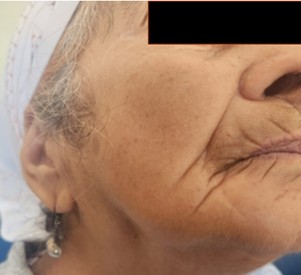
Case report: A 82 years old female patient, was referred to our department for progressive swallowing difficulties, alteration of taste sensation and mixed dysphagia and dysphonia, Without dyspnea or any associated signs. The clinical examination revealed right-sided spinal accessory nerve paresis and deviation of the soft palate to the left and a right vocal cord paralysis, these findings were consistent with a lesion involving the mixed cranial nerves. Brain MRI revealed an enlarged right jugular foramen with smooth osseous remodeling, without bone erosion or destruction. occupied by a well-defined tissue mass with marked enhancement after contrast administration. The patient was discussed at a staff meeting where a diagnosis of schwannoma involving the mixed cranial nerves was established. And Gamma Knife radiosurgery was recommended as the therapeutic approach.
Conclusion: The main points emphasize that imaging plays a crucial role in the diagnosis of jugular foramen schwannomas and can alone allow differential diagnosis from paragangliomas. Schwannomas typically appear as homogeneous soft tissue masses with regular borders, centered on the jugular foramen, and may extend to the cerebellopontine angle. They show no signs of bone destruction and lack the characteristic “salt-and-pepper” appearance seen in paragangliomas.
Downloads
References
1. Nagamine W., Conneely M., Petruzzelli G., Hacein-Bey L. Schwannome glossopharyngien de l'espace carotidien supra-hyoïdien : présentation de cas et discussion des rapports avec l'artère carotide. 2009;119(4):653–656.
2. Sweasey TA, Edelstein SR, Hoff JT. Glossopharyngeal schwannoma: review of five cases and the literature. Surg Neurol. 1991;35(2):127–130. doi:10.1016/0090-3019(91)90263-9.
3. Zhao H., Li X., Lv Q., et al. A large dumbbell-shaped glossopharyngeal schwannoma invading the vagus nerve: case report and literature review. J Med Case Rep. 2008;2:334. doi:10.1186/1752-1947-2-334.
4. Hilton DA, Hanemann CO. Schwannomas and their pathogenesis. Brain Pathol. 2014;24(3):205–220. doi:10.1111/bpa.12125.
5. Samii M, Babu RP, Tatagiba M, Sepehrnia A. Surgical treatment of jugular foramen schwannomas. J Neurosurg. 1995;82:924–932.
6. Samii M, Babu RP, Tatagiba M, Sepehrnia A. Surgical treatment of jugular foramen schwannomas. J Neurosurg. 1995;82:924–932.
7. Samii M, Babu RP, Tatagiba M, Sepehrnia A. Surgical treatment of jugular foramen schwannomas. J Neurosurg. 1995;82:924–932.
8. Gonen O., et al. Differentiation of Jugular Foramen Paragangliomas versus Schwannomas Using Golden-Angle Radial Sparse Parallel Dynamic Contrast-Enhanced MRI. AJNR. 2021;42(10):1847–1852. doi:10.3174/ajnr.A7243.
9. Guss ZD, Batra S, Li G, et al. Radiotherapy of glomus jugulare tumors: history and recent progress. Neurosurg Focus. 2009;27:E5. doi:10.3171/2009.9.FOCUS09195.